PMP
under the microscope
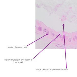
In pseudomyxoma peritonei (PMP), the cancer cells grow in the abdominal cavity where they produce large amounts of mucin.
The image shows a typical example stained with haematoxylin and eosin under the microscope.
References
The pathology of pseudomyxoma peritonei and appendix tumours
Pseudomyxoma Survivor and N Carr, MB BS FRCPath FRCPA FAcadMEd, Pseudomyxoma Survivor website, 2023
A Consensus for Classification and Pathologic Reporting of Pseudomyxoma Peritonei and Associated Appendiceal Neoplasia
Carr, Norman J. FRCPath; Cecil, Thomas D. MD; Mohamed, Faheez; Sobin, Leslie H.; Sugarbaker, Paul H.; González-Moreno, Santiago MD PhD; Taflampas, Panos MD; Chapman, Sara PhD; Moran, Brendan J. MD, A Consensus for Classification and Pathologic Reporting of Pseudomyxoma Peritonei and Associated Appendiceal Neoplasia, The American Journal of Surgical Pathology: January 2016 – Volume 40 – Issue 1 – p 14-26 doi: 10.1097/PAS.0000000000000535
This Toggle Intentionally Left Blank
Complete cytoreduction for pseudomyxoma peritonei (Sugarbaker technique)
National Institute for Health and Care Excellence (NICE) Complete cytoreduction for pseudomyxoma peritonei (Sugarbaker technique), April 2004 [Online]. Available https://www.nice.org.uk/Guidance/ipg56 [Accessed February 2018]. Under review February 2018.
Pseudomyxoma peritonei
Cancer Research UK, Pseudomyxoma peritonei, 2016 [Online]. Available http://www.cancerresearchuk.org/about-cancer/pseudomyxoma-peritonei [Accessed February 2018].
Pseudomyxoma peritonei (PMP)
Macmillan, Pseudomyxoma peritonei (PMP), 2016 [Online]. Available https://www.macmillan.org.uk/information-and-support/pseudomyxoma-peritonei-pmp [Accessed February 2018].